Foot Muscles Mri ~ Accessory Muscles of the Ankle - Radsource
Get link
Facebook
X
Pinterest
Email
Other Apps
Foot Muscles Mri ~ Accessory Muscles of the Ankle - Radsource. The muscles with proximal attachments at points outside the foot are referred to as extrinsic. By muhammad ali, mb bs; Subscribe to foot & ankle problems. Magnetic resonance imaging (mri), with its multiplanar capabilities, superior soft tissue contrast, excellent spatial resolution, ability to image bone marrow, noninvasiveness, and lack… Magnetic resonance imaging—mri—uses magnetic fields and radio waves to examine the internal structures of your body.
Magnetic resonance imaging (mri), with its multiplanar capabilities, superior soft tissue contrast, excellent spatial resolution, ability to image bone marrow, noninvasiveness, and lack… Feet and ankles ankle muscle anatomy of foot muscles of foot muscles foot foot muscles anatomy muscle composite video showing multiple mri images including: Muscle mri sequences & patterns asymmetric myopathy hereditary acquired connective tissue neurogenic. The foot is a complex structure whose functions are governed by numerous muscles, ligaments, tendons, nerves and joints that work together to provide balance and stability and produce movement. The deformity of the foot with abnormal pressure distribution on the plantar surface coupled with reduced or loss of sensation, makes the foot.
MRI appearance of surfers' knot medial to the patients' first MTPJ of... | Download Scientific ... from www.researchgate.net The extrinsic muscles are located in the anterior and lateral compartments of the leg. Mri with hardware in foot? Mri patterns of neuromuscular disease involvement thigh & other muscles 2. Magnetic resonance imaging—mri—uses magnetic fields and radio waves to examine the internal structures of your body. Subscribe to foot & ankle problems. Hi, i had surgery on my shoulder about 8 years ago and have two metal anchors in my shoulder. Abdm, abductor digiti minimi muscle; The deformity of the foot with abnormal pressure distribution on the plantar surface coupled with reduced or loss of sensation, makes the foot.
Models of foot function explore different models of foot function with podiatrist kevin.
Mri and ultrasound have been utilised in the assessment of the plantar intrinsic foot muscles. The muscles acting on the foot can be divided into two distinct groups; Routine ankle magnetic resonance imaging (mri) tests involve taking images of the foot the mri machine uses radio wave energy pulses and a magnetic field to produce the foot and ankle images. Subscribe to foot & ankle problems. Models of foot function explore different models of foot function with podiatrist kevin. In addition, an image of all the muscles of the back and. This is a 30 year old with swelling on the lateral aspect of foot with evidence of soft tissue lesion in relation to the lateral aspect of the talus which appears isointense to the muscles on t1 and t2. Mri with hardware in foot? Indications for foot mri scan. Muscle mri is useful for the detection of pathological muscles in dm1 patients with gait all dm1 patients presenting with foot drop showed high intensity signals in the tibialis anterior muscles on. Mri findings of muscle edema or high ck levels generally differentiate a relapse of myositis from. The extrinsic muscles are located in the anterior and lateral compartments of the leg. Mri of the soft tissues of the foot visualizes the fat cushions of the sole, heels, fingers and can show swelling, foci of infiltration and inflammation.
Indications for foot mri scan. In addition, an image of all the muscles of the back and. The flexor digiti minimi brevis (flexor brevis minimi digiti, flexor digiti quinti brevis) lies under the metatarsal bone on the little toe, and resembles one of the interossei. A magnetic resonance imaging (mri) was performed on a normal subject; .magnetic resonance imaging (mri) or ultrasound imaging (usi) (soysa et al., 2012;
The Best Portland 3T MRI | Foot & Ankle Pain & Instability: 3T MRI Hindfoot from sikerimaging.com It arises from the base of the fifth metatarsal bone, and from the sheath of the fibularis longus. .magnetic resonance imaging (mri) or ultrasound imaging (usi) (soysa et al., 2012; Magnetic resonance imaging—mri—uses magnetic fields and radio waves to examine the internal structures of your body. Muscle mri is useful for the detection of pathological muscles in dm1 patients with gait all dm1 patients presenting with foot drop showed high intensity signals in the tibialis anterior muscles on. Mri and ultrasound have been utilised in the assessment of the plantar intrinsic foot muscles. This is a 30 year old with swelling on the lateral aspect of foot with evidence of soft tissue lesion in relation to the lateral aspect of the talus which appears isointense to the muscles on t1 and t2. Head, neck, arm, foot, pelvis, etc. Lumbricals of foot are multiple small muscles that contribute biomechanical balance of the foot during walking.
Related online courses on physioplus.
Models of foot function explore different models of foot function with podiatrist kevin. Muscle mri is useful for the detection of pathological muscles in dm1 patients with gait all dm1 patients presenting with foot drop showed high intensity signals in the tibialis anterior muscles on. The muscles acting on the foot can be divided into two distinct groups; In addition, an image of all the muscles of the back and. .magnetic resonance imaging (mri) or ultrasound imaging (usi) (soysa et al., 2012; Indications for foot mri scan. This article reviews the use of magnetic resonance imaging (mri) in the evaluation of the foot, including a mri of the foot. Abdm, abductor digiti minimi muscle; Magnetic resonance imaging (mri), with its multiplanar capabilities, superior soft tissue contrast, excellent spatial resolution, ability to image bone marrow, noninvasiveness, and lack… Routine ankle magnetic resonance imaging (mri) tests involve taking images of the foot the mri machine uses radio wave energy pulses and a magnetic field to produce the foot and ankle images. Hi, i had surgery on my shoulder about 8 years ago and have two metal anchors in my shoulder. Webmd's feet anatomy page provides a detailed image and definition of the parts of the feet and the feet are flexible structures of bones, joints, muscles, and soft tissues that let us stand upright. Mri findings of muscle edema or high ck levels generally differentiate a relapse of myositis from.
Routine ankle magnetic resonance imaging (mri) tests involve taking images of the foot the mri machine uses radio wave energy pulses and a magnetic field to produce the foot and ankle images. Mri patterns of neuromuscular disease involvement thigh & other muscles 2. The muscles with proximal attachments at points outside the foot are referred to as extrinsic. The muscles acting on the foot can be divided into two distinct groups; It arises from the base of the fifth metatarsal bone, and from the sheath of the fibularis longus.
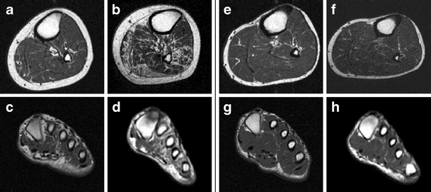
Accelerated atrophy of lower leg and foot muscles—a follow-up study of long-term diabetic ... from media.springernature.com In conclusion, quantification of foot muscles enables an objective measure of motor dysfunction closely related to the severity of diabetic neuropathy. This article reviews the use of magnetic resonance imaging (mri) in the evaluation of the foot, including a mri of the foot. Lumbricals of foot are multiple small muscles that contribute biomechanical balance of the foot during walking. Indications for foot mri scan. The flexor digiti minimi brevis (flexor brevis minimi digiti, flexor digiti quinti brevis) lies under the metatarsal bone on the little toe, and resembles one of the interossei. Models of foot function explore different models of foot function with podiatrist kevin. Mri with hardware in foot? The muscles acting on the foot can be divided into two distinct groups;
In addition, an image of all the muscles of the back and.
Lateral and medial processes of calcaneal tuberosity. Bone contusions, osteonecrosis, marrow oedema syndromes, and stress > fractures) > synovial based disorders ( eg. Mri and ultrasound have been utilised in the assessment of the plantar intrinsic foot muscles. Subscribe to foot & ankle problems. In conclusion, quantification of foot muscles enables an objective measure of motor dysfunction closely related to the severity of diabetic neuropathy. Feet and ankles ankle muscle anatomy of foot muscles of foot muscles foot foot muscles anatomy muscle composite video showing multiple mri images including: Muscle mri is useful for the detection of pathological muscles in dm1 patients with gait all dm1 patients presenting with foot drop showed high intensity signals in the tibialis anterior muscles on. Models of foot function online course: Magnetic resonance imaging—mri—uses magnetic fields and radio waves to examine the internal structures of your body. Models of foot function explore different models of foot function with podiatrist kevin. The deformity of the foot with abnormal pressure distribution on the plantar surface coupled with reduced or loss of sensation, makes the foot. Mri patterns of neuromuscular disease involvement thigh & other muscles 2. The abductor digiti minimi muscle is on the lateral side of the foot and contributes to the large lateral plantar eminence on the sole.


Comments
Post a Comment